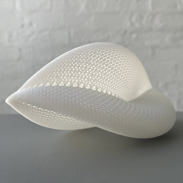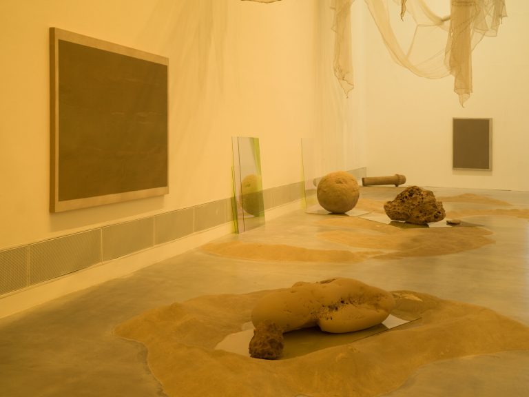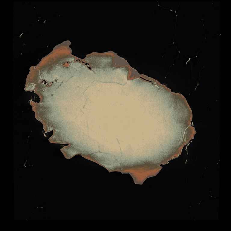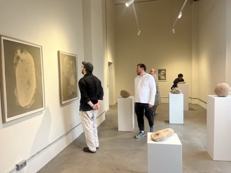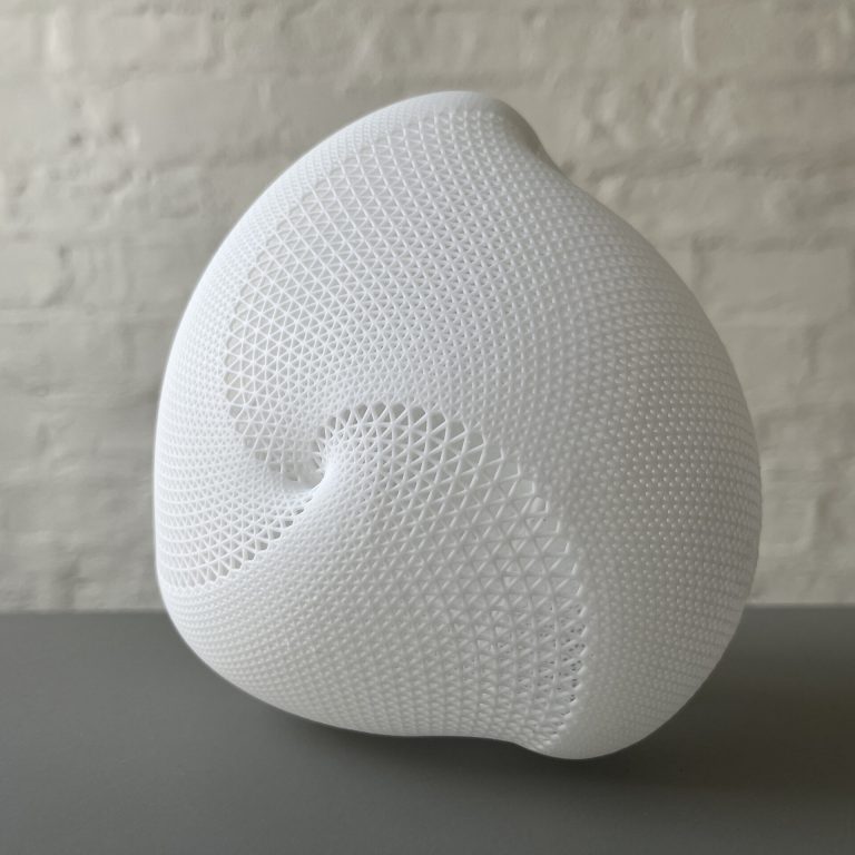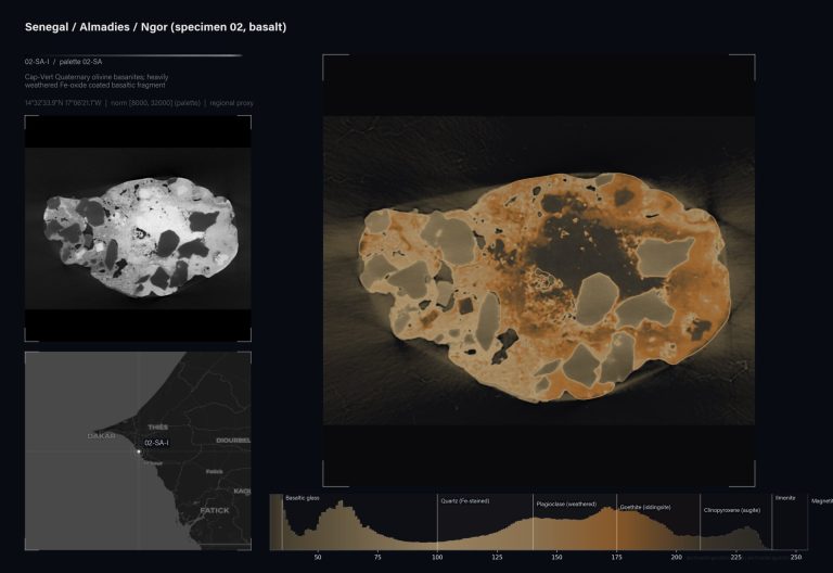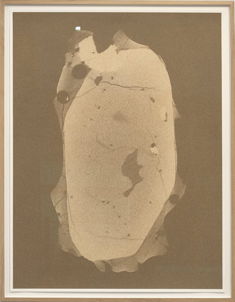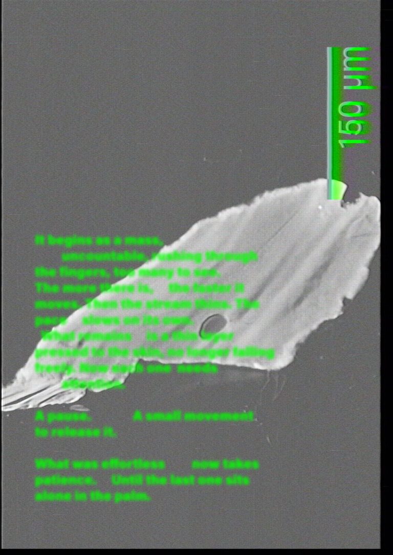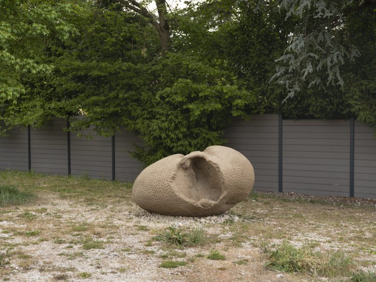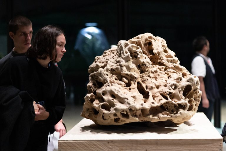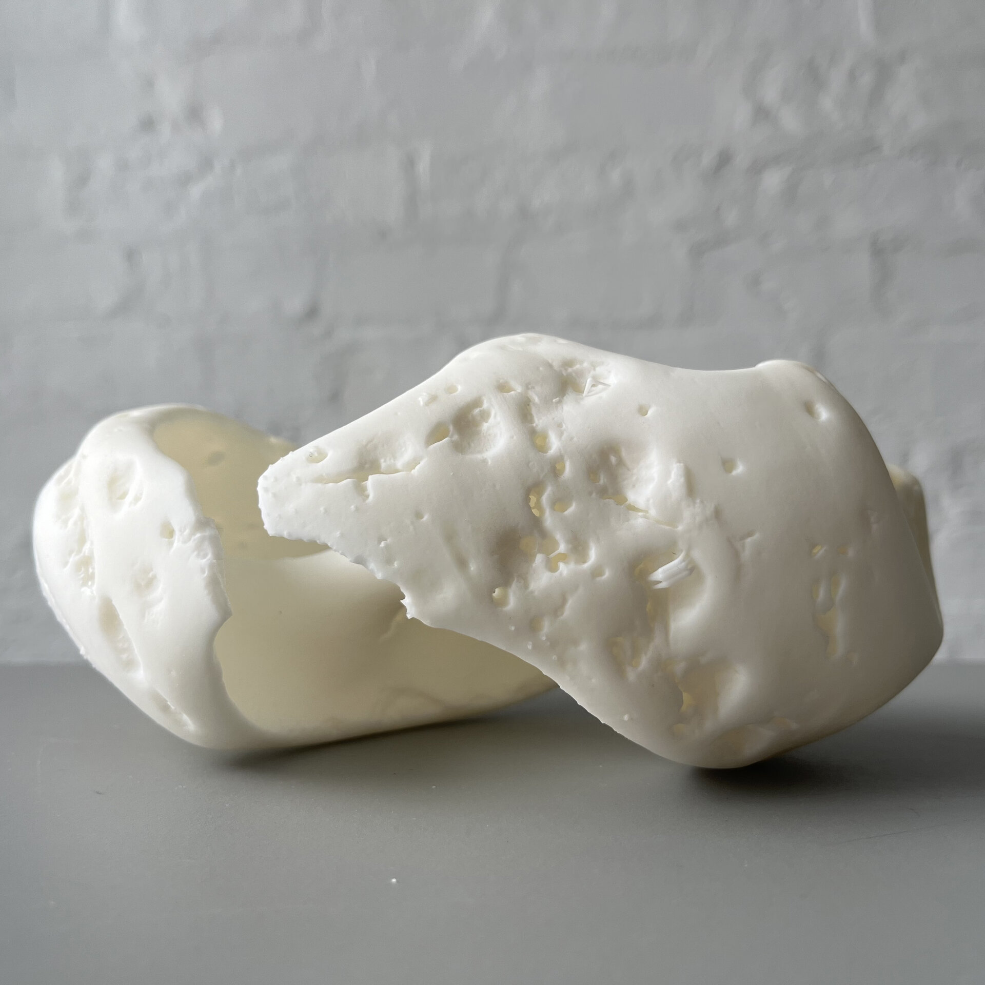
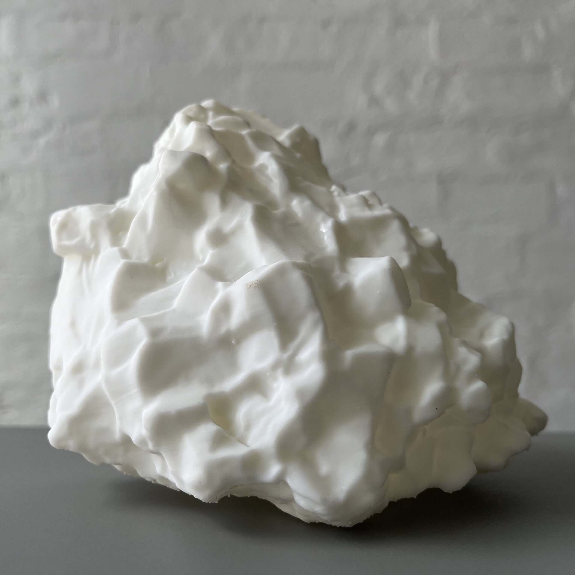
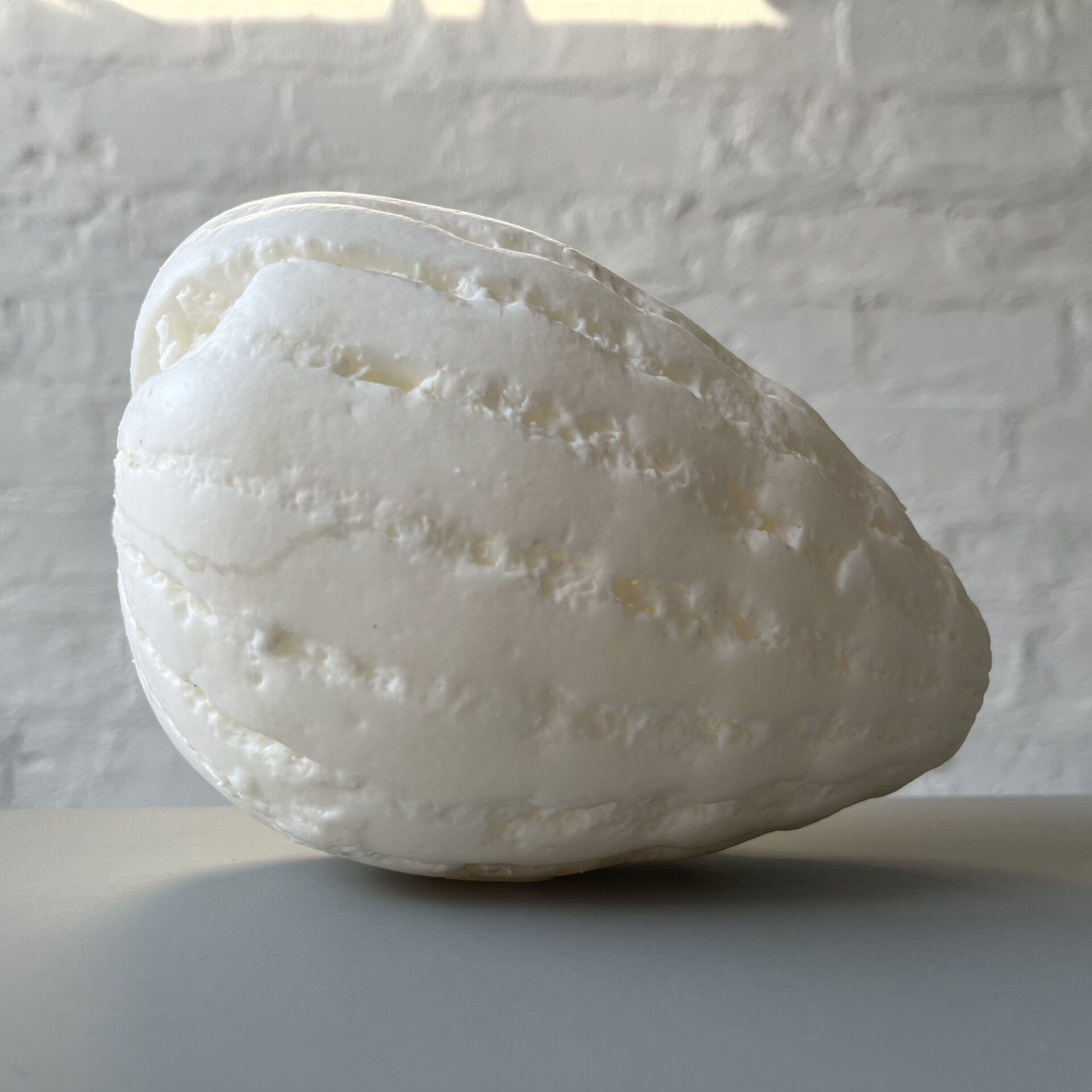
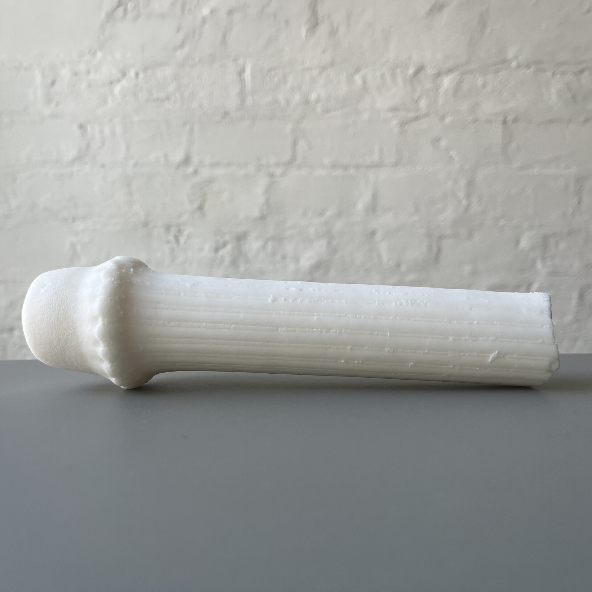
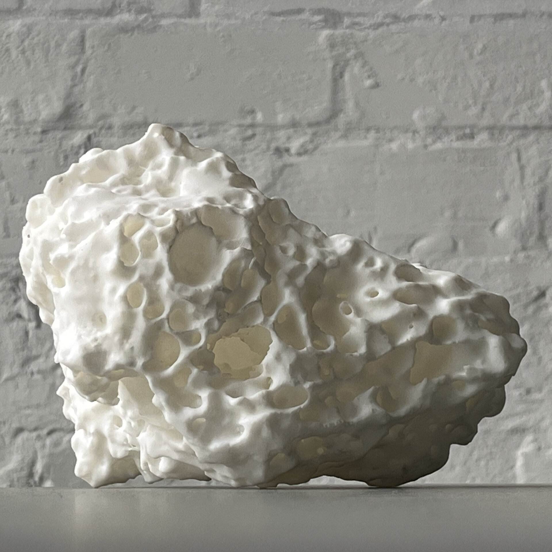
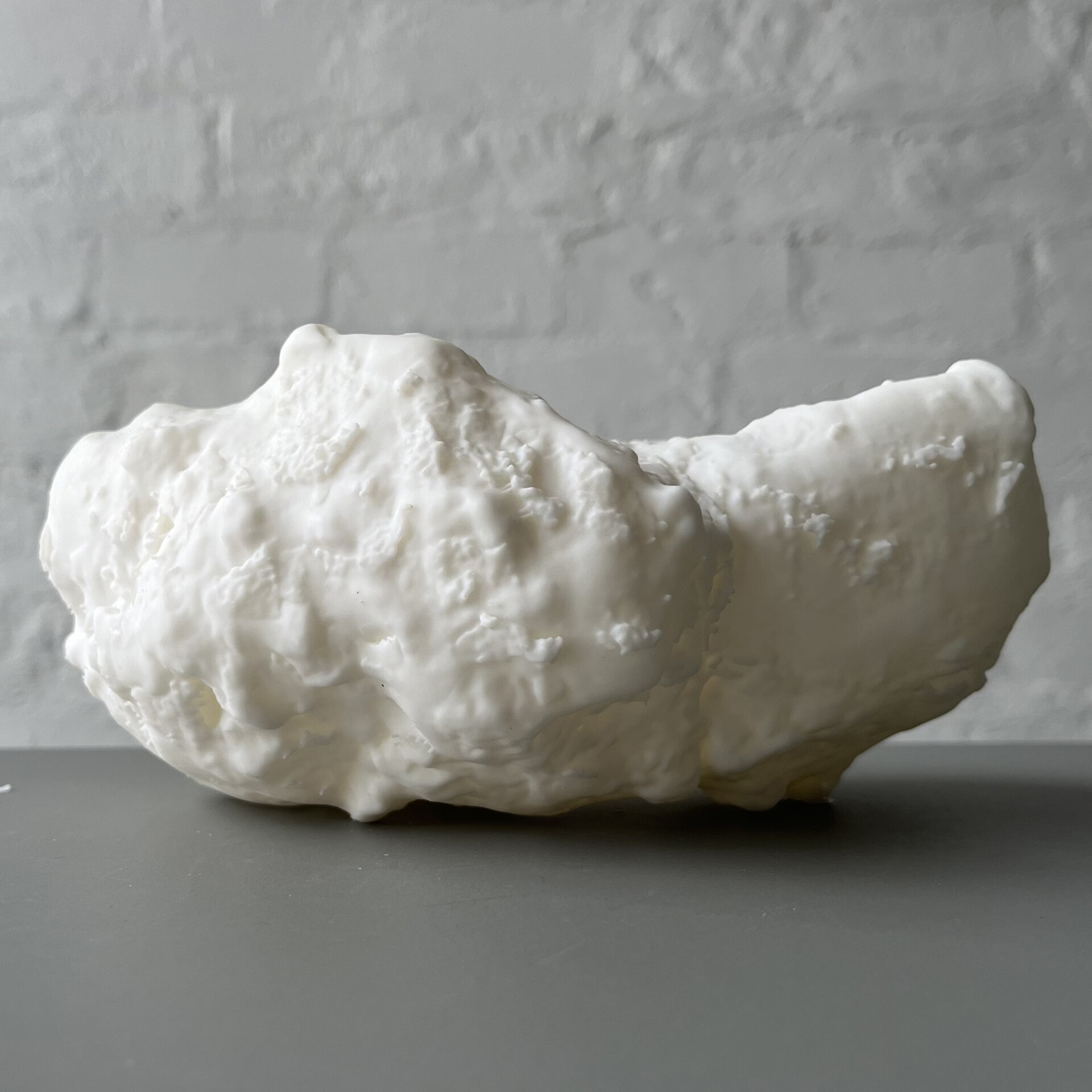
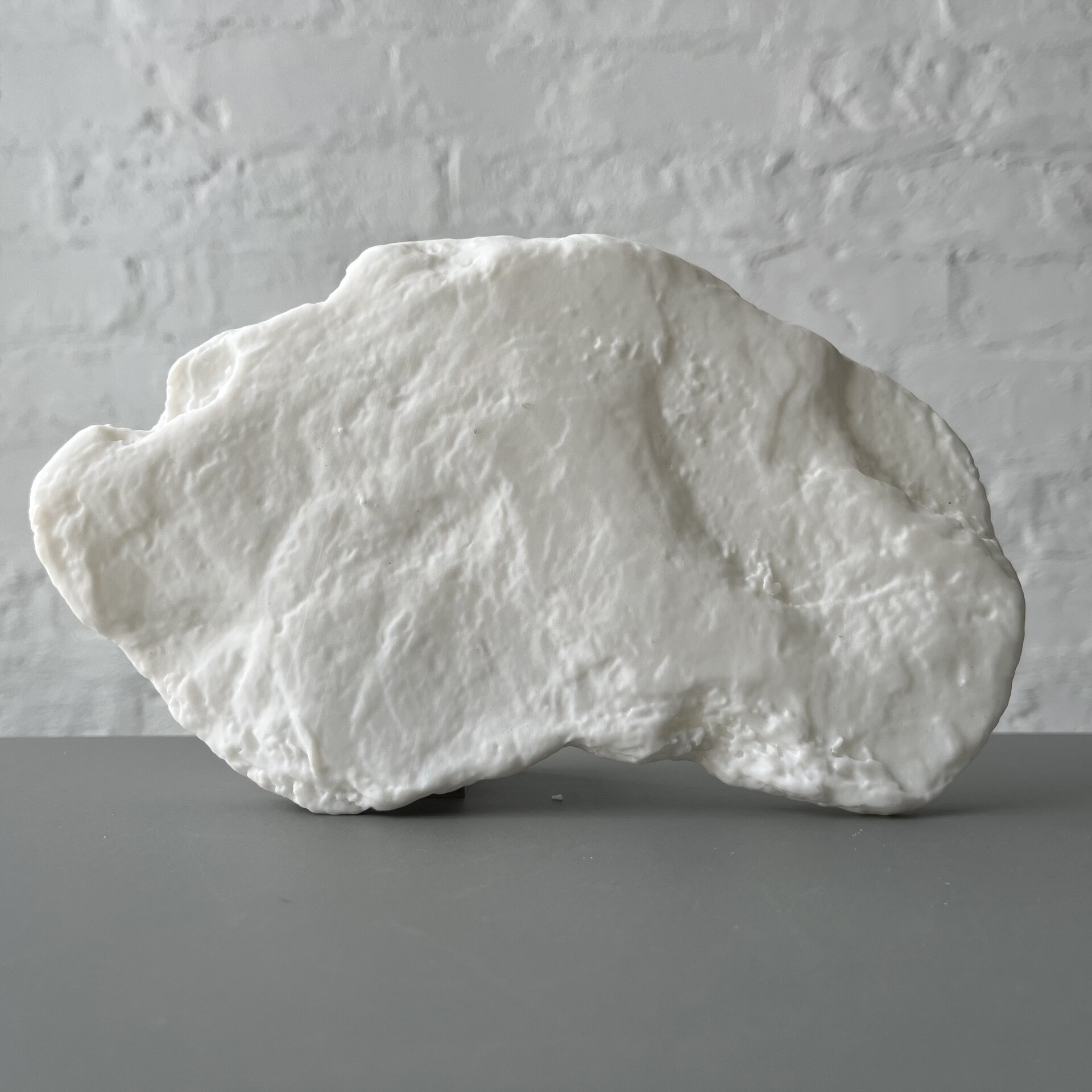
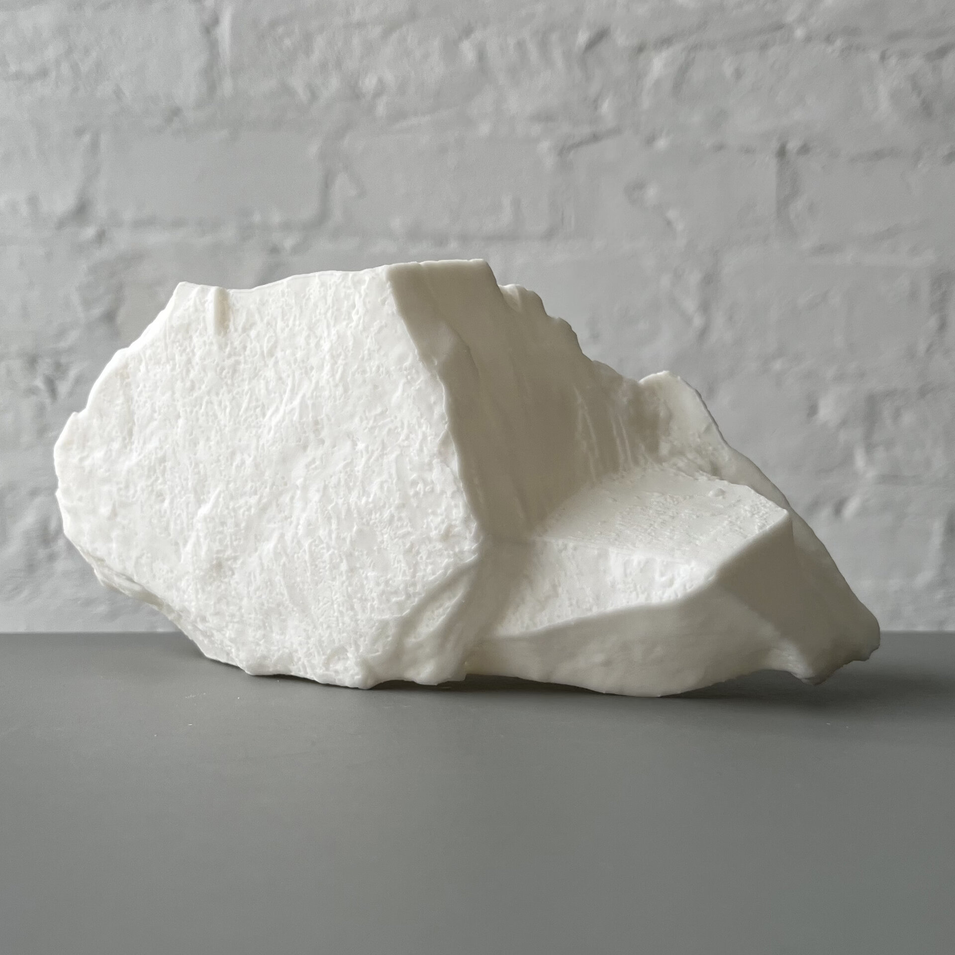
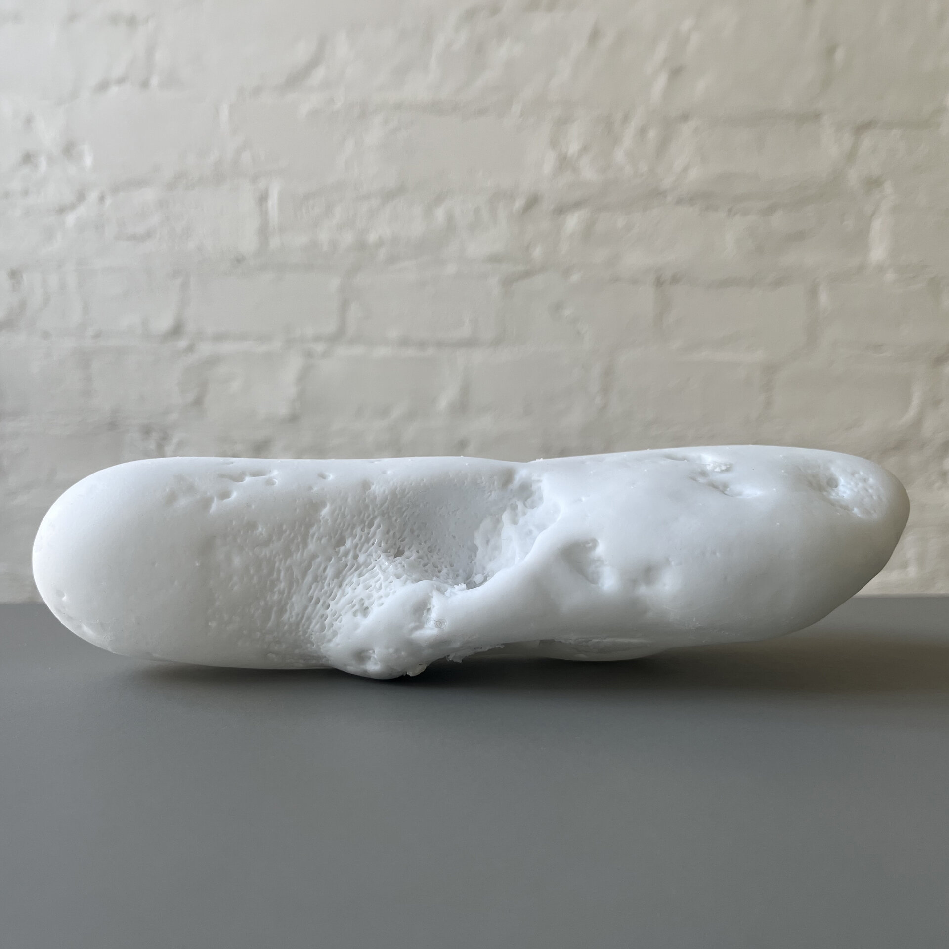
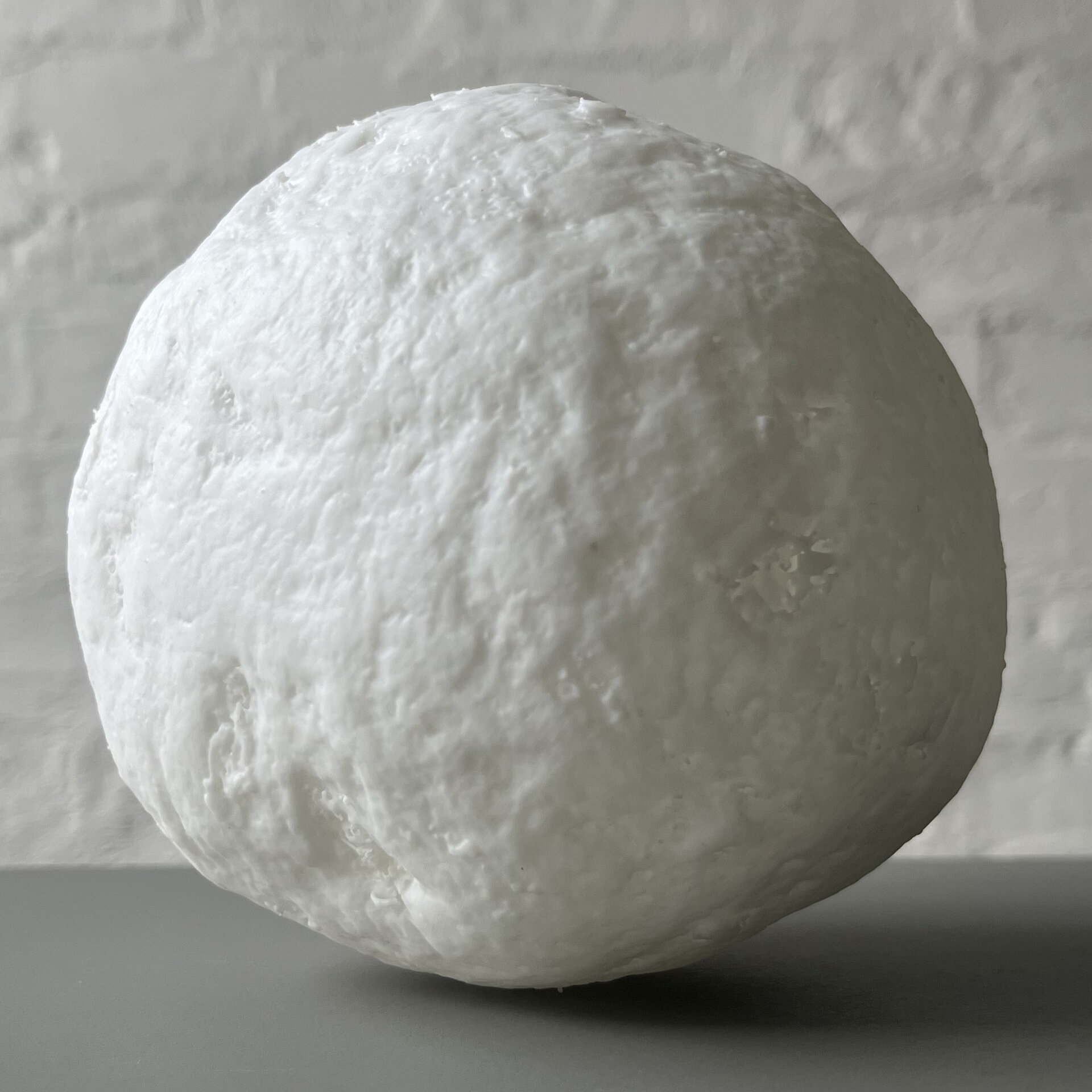
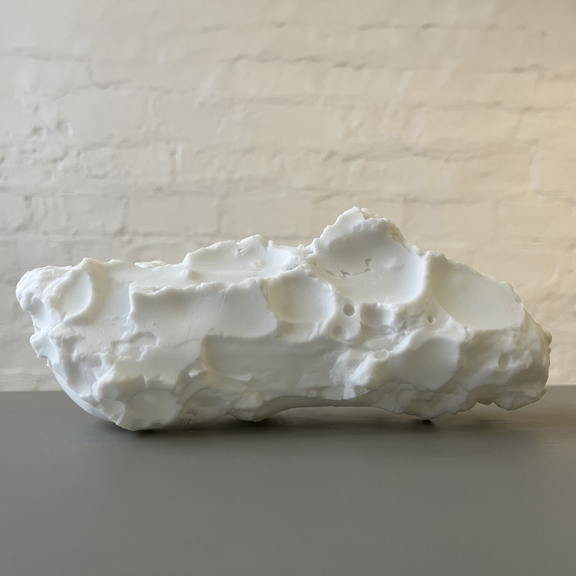
We are testing scan parameters for nano-CT imaging of sand grains. The Zeiss x-ray microscope scans the sand grains and records the data in the ubiquitous DICOM format. The images contain the microscopic surface topology information that cannot be observed by human eye. These data can be converted to buildable 3-dimensional meshes through a computational process. Sand grains carry geological information in their microscopic surface topography. The angular grains are likely to be recently broken and round grains are likely to have been transported by water for thousands of years. Pits on a sand grain surface show that it has undergone chemical weathering. We are testing different processing algorithms to see how much of the surface details can we keep with minimum wall thickness and overhang angle constraints. We are adapting medical imaging technology to geological material. Scan parameters, reconstruction algorithms, mesh generation techniques that we are researching and developing will become our protocol to capture the natural complexity at these scales where microscopic form preserves material history. This is an ongoing work.
Photography: Phillip C. Reiner
Artwork year: | Last updated: